Monthly Archives: October 2012

Scientists Identify New Stem Cells with Therapeutic Potential

 The discovery, published in the journal PLOS Biology, offers new opportunities in the treatment of cardiovascular diseases, cancer and many other diseases.

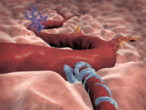
The growth of new blood vessels – angiogenesis – occurs during the repair of damaged tissue and organs in adults. However, malignant tumors also grow new blood vessels in order to receive oxygen and nutrients. As such, angiogenesis is both beneficial and detrimental to health, depending on the context, requiring therapeutic approaches that can either help to stimulate or prevent it. Therapeutics that aim to prevent the growth of new blood vessels are already in use, but the results are often more modest than predicted.

For more than a decade, Prof Petri Salvén of the University of Helsinki and his colleagues have studied the mechanisms of angiogenesis to discover how blood vessel growth could be prevented or accelerated effectively.

“We succeeded in isolating endothelial cells with a high rate of division in the blood vessel walls of mice. We found these same cells in human blood vessels and blood vessels growing in malignant tumors in humans. These cells are known as vascular endothelial stem cells. In a cell culture, one such cell is capable of producing tens of millions of new blood vessel wall cells,” Prof Salvén said.

From their studies in mice, the team was able to show that the growth of new blood vessels weakens, and the growth of malignant tumors slows, if the amount of these cells is below normal. Conversely, new blood vessels form where these stem cells are implanted.

“The identification and isolation of an entirely new adult stem cell type is a significant discovery in stem cell biology. Endothelial stem cells in blood vessels are particularly interesting, because they offer great potential for applications in practical medicine and the treatment of patients,” Prof Salvén said.

“If an efficient method of vascular endothelial stem cell production could be developed, it could offer new treatment opportunities in situations where damaged tissue or diseases call for new blood vessel growth, or where the constriction or dysfunction of blood vessels deprives tissues of oxygen, for example in cardiac disease. These cells also offer new opportunities for developing therapeutics that seek to prevent new blood vessel growth in malignant tumors.”

Cord blood studied to reverse cerebral palsy

Jenny and JD Stephenson banked their baby Weston’s umbilical cord blood when he was born.

“We weren’t guaranteed to need it for anything,” said Jenny Stephenson. 

In retrospect they are glad they had banked it. When Weston was ten months old doctors diagnosed him with cerebral palsy. It was devastating for his mother.

“Of course you want the best for him, and you never want anything to be wrong,” said Stephenson.

Weston is developmentally delayed and has trouble using his left side. There’s no cure but now there’s hope, from the toddler’s own cord blood. As part of a research study Weston recently got his first infusion.

Duke University Medical Center’s Dr. Joanne Kurtzberg is lead researcher in the new study.

“If this is beneficial, it could really change the lives of those children,” said Dr. Kurtzberg.

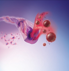
Dr. Kurtzberg said the theory is that cord blood cells can coax normal cells into fixing damaged tissue, and grow into new cells.

Cord blood cells can graft and grow into some types of brain cells,” she said.

Patients in an earlier study reported improved speech, mobility, and movement. But that study didn’t compare cord blood to a placebo. The new study does, offering hope to a little boy whose parents want the best for him.

“It’s why you get up every day and go to work,” said J.D. Stephanson, Weston’s father.

“My hope is that we see a miracle, really,” said Jennifer Stephenson.

If successful, the treatment has the potential of being that miracle for Weston and other children with cerebral palsy.

The researchers are still looking for families of kids six and under who have cerebral palsy to participate.

Source: KING 5 HealthLink


Stem Cells Show Early Promise for Rare Brain Disorder

Four young boys with a rare, fatal brain condition have made it through a dangerous ordeal. Scientists have safely transplanted human neural stem cells into their brains. Twelve months after the surgeries, the boys have more myelin — a fatty insulating protein that coats nerve fibers and speeds up electric signals between neurons — and show improved brain function, a new study in Science Translational Medicine reports. The preliminary trial paves the way for future research into potential stem cell treatments for the disorder, which overlaps with more common diseases such as Parkinson’s disease and multiple sclerosis. 

“This is very exciting,” says Douglas Fields, a neuroscientist at the National Institutes of Health in Bethesda, Maryland, who was not involved in the work. “From these early studies one sees the promise of cell transplant therapy in overcoming disease and relieving suffering.”

Without myelin, electrical impulses traveling along nerve fibers in the brain can’t travel from neuron to neuron says Nalin Gupta, lead author of the study and a neurosurgeon at the University of California, San Francisco (UCSF). Signals in the brain become scattered and disorganized, he says, comparing them to a pile of lumber. “You wouldn’t expect lumber to assemble itself into a house,” he notes, yet neurons in a newborn baby’s brain perform a similar feat with the help of myelin-producing cells called oligodendrocytes. Most infants are born with very little myelin and develop it over time. In children with early-onset Pelizaeus-Merzbacher disease, he says, a genetic mutation prevents oligodendrocytes from producing myelin, causing electrical signals to die out before they reach their destinations. This results in serious developmental setbacks, such as the inability to talk, walk, or breathe independently, and ultimately causes premature death.

Led by Gupta, the researchers drilled four small holes in each child’s skull and then used a fine needle to insert millions of stem cells into white matter deep in their frontal lobes. The scientists administered a drug that suppressed the boys’ immune systems for 9 months to keep them from rejecting the cells and checked their progress with magnetic resonance imaging and a variety of psychological and motor tests. After a year, each of the boys showed brain changes consistent with increased myelination and no serious side effects such as tumors, says David Rowitch, one of the neuroscientists on the UCSF team. In addition, three of the four boys showed “modest” improvements in their development. For example, the 5-year-old boy — the oldest child in the study — had begun for the first time to feed himself and walk with minimal assistance.

Although these signs are encouraging, Gupta and Rowitch say, a cure for Pelizaeus-Merzbacher disease is not near. Animal studies strongly support the idea that the stem cells are producing myelin-making oligodendrocytes in the boys, but it’s possible that the myelination didn’t result from the transplant but from a bout of normal growth. Rowitch adds that although such behavioral improvements are unusual for the disease, they could be a fluke. Huhn acknowledges that the study is small and has no control, but he’s is still excited. “We are for the first time seeing a biological effect of a neural stem cells transplantation into the brain [in humans].” The most important thing, he says, is that the transplants appear safe. This gives the researchers a green light to pursue larger, controlled studies, he says.

It “isn’t the flashiest thing,” but demonstrating that it’s feasible to transplant these stem cells into children’s brains without negative consequences — at least so far — is “extremely hopeful,” says Timothy Kennedy, a neuroscientist at McGill University in Montreal, Canada.

Although he’s concerned that myelination seen in mouse models might not “scale up” to a disease as severe as Pelizaeus-Merzbacher in humans, Ian Duncan, a neuroscientist at the University of Wisconsin, Madison, describes the study as setting a precedent for translating animal research in stem cells to humans. If you could improve quality of life by targeting key areas of the brain with these cells, he says, “that would be a huge advance.”


Source: ScienceNow, Wired Science


Blind Mice Get Experimental Stem Cell Treatment For Blindness

 Columbia University ophthalmologists and stem cell researchers have developed an experimental treatment for blindness using the patient’s skin cells, which has improved the vision of blind mice in testing.

The findings of this research, published online in the journal Molecular Medicine, suggest that induced pluripotent stem cells (iPS) could soon be used to improve vision in people with macular degeneration and other eye retina diseases.

“With eye diseases, I think we’re getting close to a scenario where a patient’s own skin cells are used to replace retina cells destroyed by disease or degeneration,” says Stephen Tsang, MD, PhD, associate professor of ophthalmology and pathology & cell biology. “It’s often said that iPS transplantation will be important in the practice of medicine in some distant future, but our paper suggests the future is almost here.”

Scientists were very excited by the advent of human iPS cells when they were discovered in 2007, as they provide a way to avoid the ethical complications of embryonic stem cells. Another advantage is that the iPS cells are created from the patient’s own skin, eliminating the need for anti-rejection medications. Like the ethically challenged embryonic cells, iPS cells can develop into any type of cell. To-date, no iPS cells have been implanted into people, but many ophthalmologists say that the eye would prove to be ideal testing ground for iPS therapies.

“The eye is a transparent and accessible part of the central nervous system, and that’s a big advantage. We can put cells into the eye and monitor them every day with routine non-invasive clinical exams,” Tsang said. “And in the event of serious complications, removing the eye is not a life-threatening event.”

Professor Tsang is running a new preclinical iPS study using human iPS cells derived from the skin cells of a 53-year-old donor. The cells were first transformed with a cocktail of growth factors into cells in the retina that lie underneath the eye’s light-sensing cells.

Retina cells nourish the light-sensing cells and protect the fragile cells from excess light, heat and cellular debris. In macular degeneration and retinitis pigmentosa, retina cells die, which allows the photoreceptor cells to degenerate causing the patient to lose their vision. It is estimated that 30 percent of people will have some form of macular degeneration by the time they are 75 years old, as it is the leading cause of vision loss in the elderly. Currently, it affects 7 million Americans and that is expected to double by 2020.

The Columbia research team injected the iPS-derived retina cells into the right eyes of 34 mice that had a genetic mutation that caused their retina cells to degenerate. In many of the mice, the iPS cells assimilated into the retina without disruption and functioned as normal retina cells well into the animal’s old age.  Mice in the control group, who received injections of saline or inactive cells, showed no improvement in retina tests.

“Our findings provide the first evidence of life-long neuronal recovery in a preclinical model of retinal degeneration, using stem cell transplant, with vision improvement persisting through the lifespan,” Tsang says. “And importantly, we saw no tumors in any of the mice, which should allay one of the biggest fears people have about stem cell transplants: that they will generate tumors.”

They hope to begin a clinical trial for macular degeneration patients in the next three years, but first need to complete more preclinical testing in animal models.

“We have a good idea which patients will eventually lose their vision. In the early stages of macular degeneration we can tell by looking in the eye, and new genetic tests can now predict vision loss with 70 percent accuracy even before those signs emerge,” Tsang says. “If the therapy is safe, we could intervene very early to prevent much vision loss.”

Source: April Flowers, RedOrbit.com